
Evaluation of Functionality and Biological Responses of Mytilus galloprovincialis after Exposure to Quaternium-15 (Methenamine 3-Chloroallylochloride)


 ,
,  , , and
Abstract
:1. Introduction
2. Results
2.1. Monitoring Parameters
2.2. Histopathological Condition Indices

| Histopathological Parameters | Alteration | w | 1.0 mg/L | 2.0 mg/L |
|---|---|---|---|---|
| Cellular and morphological changes | 0.48 ± 0.04 | 0.74 ± 0.06 | ||
| Melanin/lipofuscin deposit | 1 | |||
| Loss of cilia | 2 | |||
| Enlarged central vessel | 1 | |||
| Haemocyte infiltration | 1 | |||
| Hypertrophy of goblet cells (GC) | 2 |
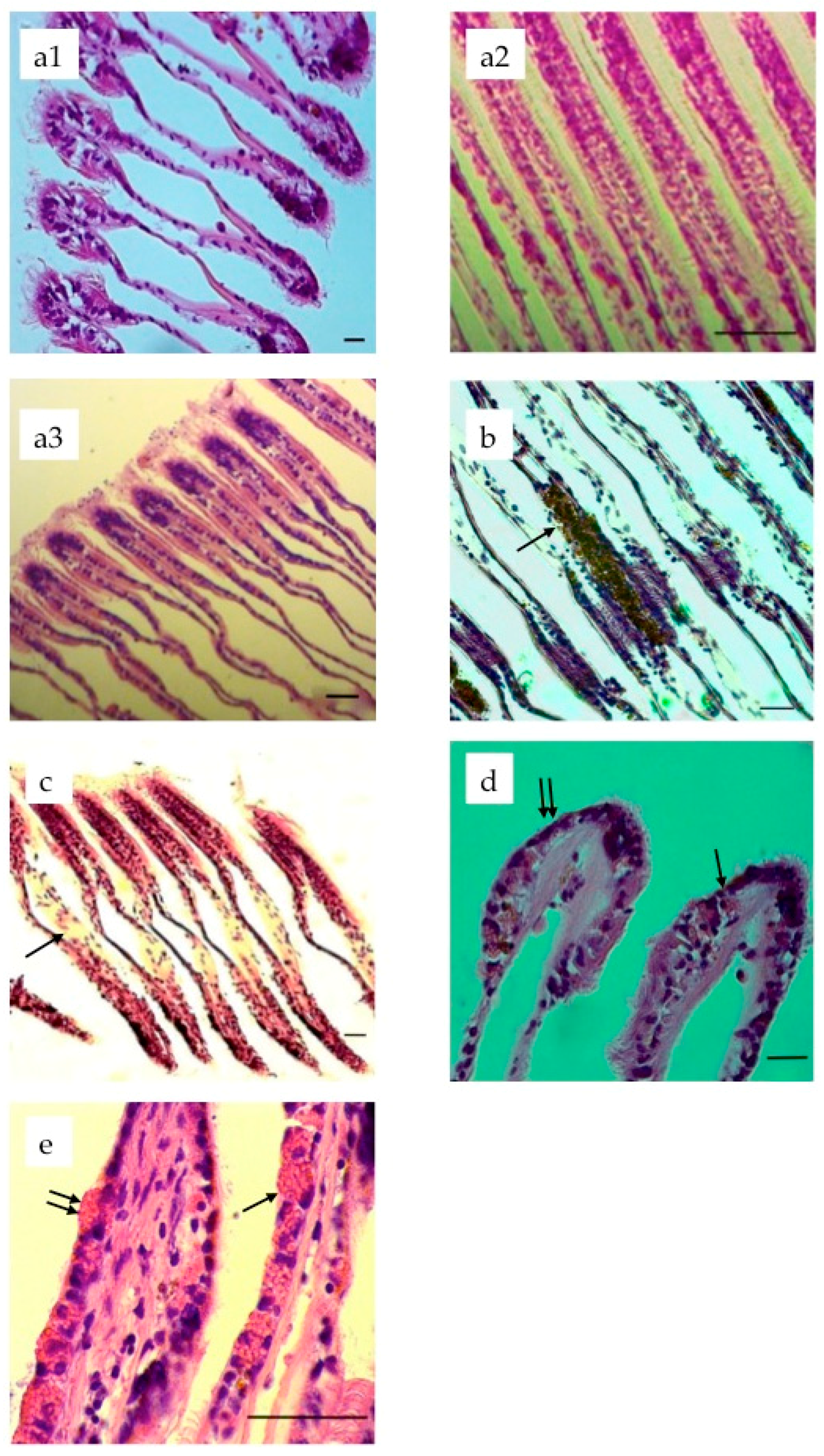
2.3. Histopathology
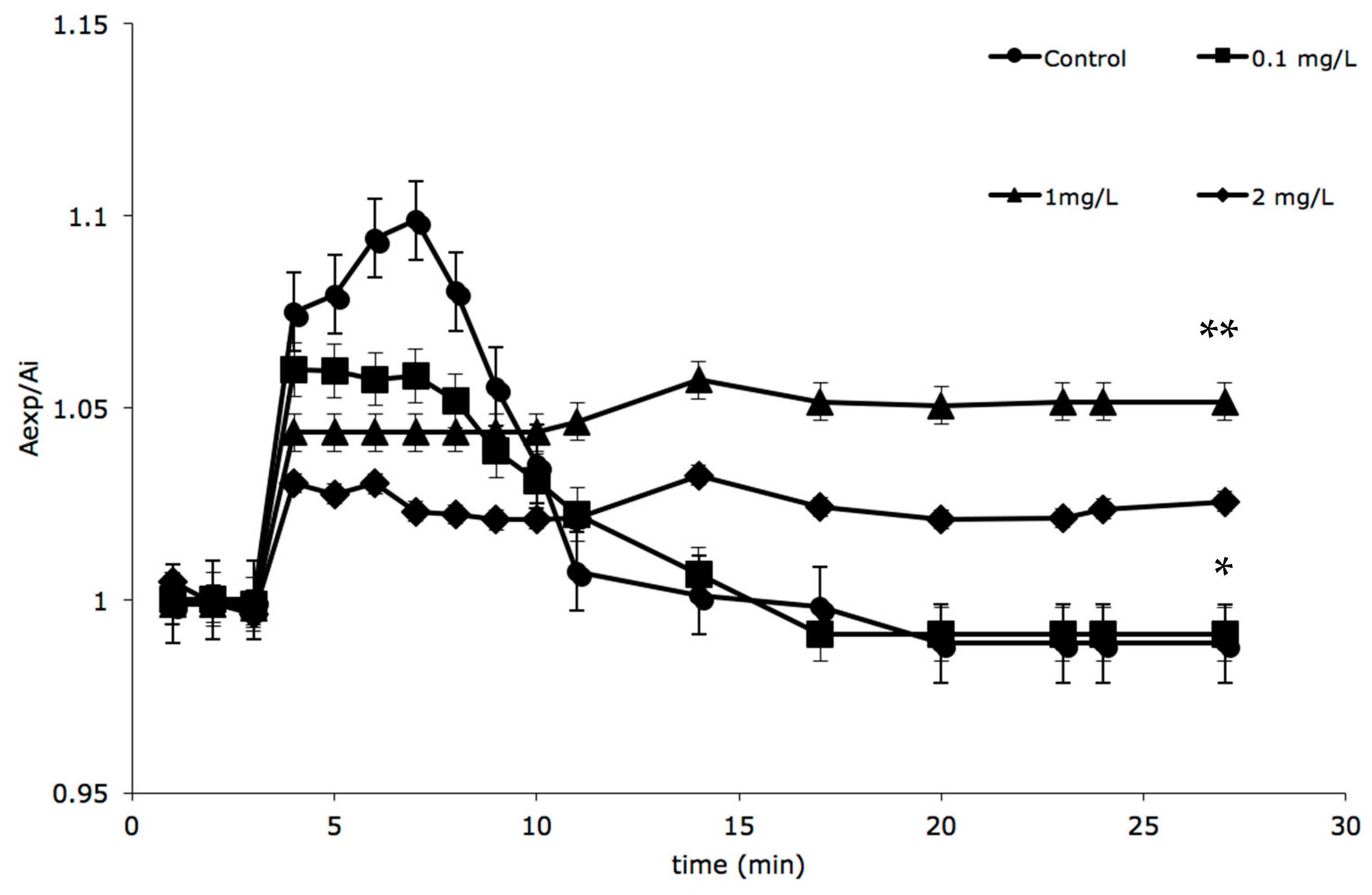
2.4. RVD Experiments in Digestive Cells


3. Discussion
4. Experimental Section
4.1. Animal Collection
4.2. Water Monitoring
4.3. Histological Analysis
4.4. Histopathological Condition Indices
4.5. Isolation of Digestive Cells and RVD Experiments
4.6. Statistical Analysis
5. Conclusions
Supplementary Materials
Acknowledgments
Author Contributions
Conflicts of Interest
References
- Wielogórska, E.; Elliott, C.T.; Danaher, M.; Connolly, L. Endocrine disruptor activity of multiple environmental food chain contaminants. Toxicol. In Vitro 2015, 29, 211–220. [Google Scholar] [CrossRef] [PubMed]
- Haman, C.; Dauchy, X.; Rosin, C.; Munoz, J.-F. Occurrence, fate and behavior of parabens in aquatic environments: A review. Water Res. 2015, 68, 1–11. [Google Scholar] [CrossRef] [PubMed]
- Brandão, F.P.; Pereira, J.L.; Gonçalves, F.; Nunes, B. The impact of paracetamol on selected biomarkers of the mollusc species Corbicula fluminea. Environ. Toxicol. 2014, 29, 74–83. [Google Scholar] [CrossRef] [PubMed]
- Lv, C.; Hou, J.; Xie, W.; Cheng, H. Investigation on formaldehyde release from preservatives in cosmetics. Int. J. Cosmet. Sci. 2015, 37, 474–478. [Google Scholar] [CrossRef] [PubMed]
- Toholka, R.; Wang, Y.S.; Tate, B.; Tam, M.; Cahill, J.; Palmer, A.; Nixon, R. The first Australian Baseline Series: Recommendations for patch testing in suspected contact dermatitis. Australas. J. Dermatol. 2014. [Google Scholar] [CrossRef] [PubMed]
- Shaughnessy, C.N.; Malajian, D.; Belsito, D.V. Cutaneous delayed-type hypersensitivity in patients with atopic dermatitis: Reactivity to topical preservatives. J. Am. Acad. Dermatol. 2014, 70, 102–107. [Google Scholar] [CrossRef] [PubMed]
- Maier, L.E.; Lampel, H.P.; Bhutani, T.; Jacob, S.E. Hand dermatitis: A focus on allergic contact dermatitis to biocides. Dermatol. Clin. 2009, 27, 251–264. [Google Scholar] [CrossRef] [PubMed]
- Marks, J.G.; Belsito, D.V.; DeLeo, V.A.; Fowler, J.F.; Fransway, A.F.; Maibach, H.I.; Mathias, C.T.; Nethercott, J.R.; Rietschel, R.L.; Sherertz, E.F. North American Contact Dermatitis Group patch test results for the detection of delayed-type hypersensitivity to topical allergens. J. Am. Acad. Dermatol. 1998, 38, 911–918. [Google Scholar] [CrossRef]
- Odhav, A.; Belsito, D.V. Is quaternium-15 a formaldehyde releaser? Correlation between positive patch test reactions to formaldehyde and quaternium-15. Dermatitis 2012, 23, 39–43. [Google Scholar] [CrossRef] [PubMed]
- Restani, P.; Galli, C.L. Oral toxicity of formaldehyde and its derivatives. CRC Crit. Rev. Toxicol. 1991, 21, 315–328. [Google Scholar] [CrossRef] [PubMed]
- Warshaw, E.M.; Ahmed, R.L.; Belsito, D.V.; DeLeo, V.A.; Fowler, J.F.; Maibach, H.I.; Marks, J.G.; Mathias, C.T.; Pratt, M.D.; Rietschel, R.L. Contact dermatitis of the hands: Cross-sectional analyses of North American Contact Dermatitis Group Data, 1994–2004. J. Am. Acad. Dermatol. 2007, 57, 301–314. [Google Scholar] [CrossRef] [PubMed]
- Siti Zulaikha, R.; Sharifah Norkhadijah, S.I.; Praveena, S.M. Hazardous ingredients in cosmetics and personal care products and health concern: A review. Public Health Res. 2015, 5, 7–15. [Google Scholar]
- Munoz, P.; Meseguer, J.; Esteban, M.Á. Phenoloxidase activity in three commercial bivalve species. Changes due to natural infestation with Perkinsus atlanticus. Fish Shellfish Immunol. 2006, 20, 12–19. [Google Scholar] [CrossRef] [PubMed]
- Messina, C.M.; Faggio, C.; Laudicella, V.A.; Sanfilippo, M.; Trischitta, F.; Santulli, A. Effect of sodium dodecyl sulfate (SDS) on stress response in the Mediterranean mussel (Mytilus Galloprovincialis): Regulatory volume decrease (Rvd) and modulation of biochemical markers related to oxidative stress. Aquat. Toxicol. 2014, 157, 94–100. [Google Scholar] [CrossRef] [PubMed]
- Matozzo, V.; Chinellato, A.; Munari, M.; Bressan, M.; Marin, M.G. Can the combination of decreased pH and increased temperature values induce oxidative stress in the clam Chamelea gallina and the mussel Mytilus galloprovincialis? Mar. Pollut. Bull. 2013, 72, 34–40. [Google Scholar] [CrossRef] [PubMed]
- Matozzo, V.; Chinellato, A.; Munari, M.; Finos, L.; Bressan, M.; Marin, M.G. First evidence of immunomodulation in bivalves under seawater acidification and increased temperature. PLoS ONE 2012, 7, e33820. [Google Scholar] [CrossRef] [PubMed]
- Marigómez, I.; Zorita, I.; Izagirre, U.; Ortiz-Zarragoitia, M.; Navarro, P.; Etxebarria, N.; Orbea, A.; Soto, M.; Cajaraville, M.P. Combined use of native and caged mussels to assess biological effects of pollution through the integrative biomarker approach. Aquat. Toxicol. 2013, 136, 32–48. [Google Scholar] [CrossRef] [PubMed]
- Krmpotić, M.; Rožmarić, M.; Barišić, D. Mussels (Mytilus galloprovincialis) as a bio-indicator species in radioactivity monitoring of Eastern Adriatic coastal waters. J. Environ. Radioact. 2015, 144, 47–51. [Google Scholar] [CrossRef] [PubMed]
- Rocha, T.L.; Gomes, T.; Cardoso, C.; Letendre, J.; Pinheiro, J.P.; Sousa, V.S.; Teixeira, M.R.; Bebianno, M.J. Immunocytotoxicity, cytogenotoxicity and genotoxicity of cadmium-based quantum dots in the marine mussel Mytilus galloprovincialis. Mar. Environ. Res. 2014, 101, 29–37. [Google Scholar] [CrossRef] [PubMed]
- Kumar, V.; Sinha, A.K.; Rodrigues, P.P.; Mubiana, V.K.; Blust, R.; de Boeck, G. Linking environmental heavy metal concentrations and salinity gradients with metal accumulation and their effects: A case study in 3 mussel species of Vitória estuary and Espírito Santo bay, Southeast Brazil. Sci. Total Environ. 2015, 523, 1–15. [Google Scholar] [CrossRef] [PubMed]
- Torre, A.; Trischitta, F.; Corsaro, C.; Mallamace, D.; Faggio, C. Digestive cells from Mytilus galloprovincialis show a partial regulatory volume decrease following acute hypotonic stress through mechanisms involving inorganic ions. Cell Biochem. Funct. 2013, 31, 489–495. [Google Scholar] [CrossRef] [PubMed]
- Resgalla, C., Jr.; Brasil, E.D.S.; Salomão, L.C. The effect of temperature and salinity on the physiological rates of the mussel Perna perna (Linnaeus 1758). Braz. Arch. Biol. Technol. 2007, 50, 543–556. [Google Scholar] [CrossRef]
- Brahim Errahmani, M.; Zouaoui, F.; Bendjoudi, D. Metabolic effects in the bivalve Perna perna and Mytilus galloprovincialis: Impact on the environment due to contamination by copper. J. Mar. Biol. 2014, 2014, Article ID 913932. [Google Scholar] [CrossRef]
- Shirayama, Y. Towards comprehensive understanding of impacts on marine organisms due to raised CO2 concentration. In Proceedings of the 5th International Symposiumon CO2 Fixation and Efficient Utilization of Energy, Tokyo, Japan, 4–6 March 2002; pp. 177–181.
- Ringwood, A.H.; Keppler, C.J. Water quality variation and clam growth: Is pH really a non-issue in estuaries? Estuaries 2002, 25, 901–907. [Google Scholar] [CrossRef]
- Michaelidis, B.; Ouzounis, C.; Paleras, A.; Pörtner, H.O. Effects of long-term moderate hypercapnia on acid-base balance and growth rate in marine mussels Mytilus galloprovincialis. Mar. Ecol. Prog. Ser. 2005, 293, 109–118. [Google Scholar] [CrossRef]
- Takeuchi, K.; Fujioka, Y.; Kawasaki, Y.; Shirayama, Y. Impacts of high concentration of CO2 on marine organisms; a modification of CO2 ocean sequestration. Energ. Convers. Manag. 1997, 38, S337–S341. [Google Scholar] [CrossRef]
- Travassos, A.R.; Claes, L.; Boey, L.; Drieghe, J.; Goossens, A. Non-fragrance allergens in specific cosmetic products. Contact Dermat. 2011, 65, 276–285. [Google Scholar] [CrossRef] [PubMed]
- Osdoit, S.; Guillet, M.H.; Guillet, G. Contact sensitization to quaternium-15 acting as a warning sign for curare allergy. Contact Dermat. 2011, 65, 120–122. [Google Scholar] [CrossRef] [PubMed]
- De Groot, A.C.; Blok, J.; Coenraads, P.J. Relationship between formaldehyde and quaternium-15 contact allergy. Influence of strength of patch test reactions. Contact Dermat. 2010, 63, 187–191. [Google Scholar] [CrossRef] [PubMed]
- Mose, A.P.; Lundov, M.D.; Zachariae, C.; Menne, T.; Veien, N.K.; Laurberg, G.; Kaaber, K.; Avnstorp, C.; Andersen, K.E.; Paulsen, E.; et al. Occupational contact dermatitis in painters: An analysis of patch test data from the Danish Contact Dermatitis Group. Contact Dermat. 2012, 67, 293–297. [Google Scholar] [CrossRef] [PubMed]
- Chow, E.T.; Avolio, A.M.; Lee, A.; Nixon, R. Frequency of positive patch test reactions to preservatives: The Australian experience. Australas. J. Dermatol. 2013, 54, 31–35. [Google Scholar] [CrossRef] [PubMed]
- Doumit, J.; Pratt, M. Comparative study of IQ-ultra and Finn Chambers test methodologies in detecting 10 common standard allergens that cause allergic contact dermatitis. J. Cutan. Med. Surg. 2012, 16, 18–22. [Google Scholar] [PubMed]
- Carew, B.; Muir, J. Patch testing for allergic contact dermatitis to cigarettes: Smoked/unsmoked components and formaldehyde factors. Australas. J. Dermatol. 2014, 55, 225–226. [Google Scholar] [CrossRef] [PubMed]
- O’Gorman, S.M.; Torgerson, R.R. Allergic contact dermatitis of the vulva. Dermatitis 2013, 24, 64–72. [Google Scholar] [CrossRef] [PubMed]
- Liu, J.; Pan, L.-Q.; Zhang, L.; Miao, J.; Wang, J. Immune responses, ROS generation and the haemocyte damage of scallop Chlamys farreri exposed to Aroclor 1254. Fish Shellfish Immunol. 2009, 26, 422–428. [Google Scholar] [CrossRef] [PubMed]
- Larguinho, M.; Cordeiro, A.; Diniz, M.S.; Costa, P.M.; Baptista, P.V. Metabolic and histopathological alterations in the marine bivalve Mytilus galloprovincialis induced by chronic exposure to acrylamide. Environ. Res. 2014, 135, 55–62. [Google Scholar] [CrossRef] [PubMed]
- Carballeira, C.; Espinosa, J.; Carballeira, A. Linking δ 15 N and histopathological effects in molluscs exposed in situ to effluents from land-based marine fish farms. Mar. Pollut. Bull. 2011, 62, 2633–2641. [Google Scholar] [CrossRef] [PubMed]
- Auffret, M. Histopathological changes related to chemical contamination in Mytilus edulis from field and experimental conditions. Mar. Ecol. Prog. Ser. 1988, 46, 101–107. [Google Scholar] [CrossRef]
- El-Shenawy, N.S.; Greenwood, R.; Abdel-Nabi, I.M. Histological responses of marine mussel; Mytilus edulis to long-term exposure to sublethal-level of lindane and atrazine. Acta Zool. Sin. 2007, 53, 899–909. [Google Scholar]
- Miao, J.-J.; Pan, L.-Q.; Liu, J.; Zhang, L. Effects of benzo[a]pyrene on DNA damage and histological alterations in gonad of scallop Chlamys farreri. Mar. Environ. Res. 2009, 67, 47–52. [Google Scholar] [CrossRef] [PubMed]
- Pan, L.; Ren, J.; Liu, J. Effects of benzo (k) fluoranthene exposure on the biomarkers of scallop Chlamys farreri. Comp. Biochem. Phys. C 2005, 141, 248–256. [Google Scholar] [CrossRef] [PubMed]
- Zuykov, M.; Pelletier, E.; Harper, D.A. Bivalve mollusks in metal pollution studies: From bioaccumulation to biomonitoring. Chemosphere 2013, 93, 201–208. [Google Scholar] [CrossRef] [PubMed]
- Antunes, S.; Freitas, R.; Figueira, E.; Gonçalves, F.; Nunes, B. Biochemical effects of acetaminophen in aquatic species: Edible clams Venerupis decussata and Venerupis philippinarum. Environ. Sci. Pollut. Res. Int. 2013, 20, 6658–6666. [Google Scholar] [CrossRef] [PubMed]
- Auriemma, R.; Battistella, S. Biochemical and histological alterations of Mytilus galloprovincialis digestive gland after exposure to okadaic acid and derivatives. ISJ 2004, 1, 66–71. [Google Scholar]
- Munari, M.; Marin, M.G.; Matozzo, V. Effects of the antidepressant fluoxetine on the immune parameters and acetylcholinesterase activity of the clam Venerupis philippinarum. Mar. Environ. Res. 2014, 94, 32–37. [Google Scholar] [CrossRef] [PubMed]
- Torre, A.; Trischitta, F.; Faggio, C. Effect of CdCl2 on Regulatory Volume Decrease (RVD) in Mytilus galloprovincialis digestive cells. Toxicol. Vitro 2013, 27, 1260–1266. [Google Scholar] [CrossRef] [PubMed]
- Dianzani, U.M.; Dianzani, I.; Dianzani, U. Istituzioni di Patologia Generale, 4th ed.; UTET Scienze Mediche: Milano, Italia, 2004; p. 720. [Google Scholar]
- De Vico, G.; Carella, F. Argomenti di Patologia Comparata dei Molluschi; Loffredo Editore: Napoli, Italy, 2012; p. 351. [Google Scholar]
- De Oliveira David, J.A.; Salaroli, R.B.; Fontanetti, C.S. The significance of changes in Mytella falcata (Orbigny, 1842) gill filaments chronically exposed to polluted environments. Micron 2008, 39, 1293–1299. [Google Scholar] [CrossRef] [PubMed]
- Carella, F. Biotechnologies to Evaluate the Environmental Status: New Test Organisms in Ecotoxicology and Histopathological and Molecular Biomarkers in Natural Population. Ph.D. Thesis, University of Naples Federico II, Naples, Italy, 2010. [Google Scholar]
- Pain, S.; Parant, M. Le mécanisme de défense multixénobiotique (MDMX) chez les bivalves. C. R. Biol. 2003, 326, 659–672. [Google Scholar] [CrossRef]
- Lambert, I.; Hoffmann, E.; Pedersen, S. Cell volume regulation: Physiology and pathophysiology. Acta Physiol. 2008, 194, 255–282. [Google Scholar] [CrossRef] [PubMed]
- Faggio, C.; Torre, A.; Pelle, E.; Raffa, F.; Villari, V.; Trischitta, F. Cell volume regulation following hypotonic shock in hepatocytes isolated from Sparus aurata. Comp. Biochem. Phys. A 2011, 158, 143–149. [Google Scholar] [CrossRef] [PubMed]
- Trischitta, F.; Denaro, M.G.; Faggio, C. Cell volume regulation following hypotonic stress in the intestine of the eel, Anguilla anguilla, is Ca2+-dependent. Comp. Biochem. Phys. B 2005, 140, 359–367. [Google Scholar] [CrossRef] [PubMed]
- Abel, P. Toxicity of synthetic detergents to fish and aquatic invertebrates. J. Fish Biol. 1974, 6, 279–298. [Google Scholar] [CrossRef]
- Bernet, D.; Schmidt, H.; Meier, W.; Burkhardt–Holm, P.; Wahli, T. Histopathology in fish: Proposal for a protocol to assess aquatic pollution. J. Fish Dis. 1999, 22, 25–34. [Google Scholar] [CrossRef]
- Costa, P.M.; Carreira, S.; Costa, M.H.; Caeiro, S. Development of histopathological indices in a commercial marine bivalve (Ruditapes decussatus) to determine environmental quality. Aquat. Toxicol. 2013, 126, 442–454. [Google Scholar] [CrossRef] [PubMed]
- Costa, P.M.; Diniz, M.S.; Caeiro, S.; Lobo, J.; Martins, M.; Ferreira, A.M.; Caetano, M.; Vale, C.; DelValls, T.Á.; Costa, M.H. Histological biomarkers in liver and gills of juvenile Solea senegalensis exposed to contaminated estuarine sediments: A weighted indices approach. Aquat. Toxicol. 2009, 92, 202–212. [Google Scholar] [CrossRef] [PubMed]
- Costa, P.M.; Caeiro, S.; Lobo, J.; Martins, M.; Ferreira, A.M.; Caetano, M.; Vale, C.; DelValls, T.Á.; Costa, M.H. Estuarine ecological risk based on hepatic histopathological indices from laboratory and in situ tested fish. Mar. Pollut. Bull. 2011, 62, 55–65. [Google Scholar] [CrossRef] [PubMed]
- Dailianis, S.; Kaloyianni, M. Cadmium induces both pyruvate kinase and Na+/H+ exchanger activity through protein kinase C mediated signal transduction, in isolated digestive gland cells of Mytilus galloprovincialis (L.). J. Exp. Biol. 2004, 207, 1665–1674. [Google Scholar] [CrossRef] [PubMed]
- Sample Availability: Samples of the compounds are not available from the authors.
© 2016 by the authors. Licensee MDPI, Basel, Switzerland. This article is an open access article distributed under the terms and conditions of the Creative Commons by Attribution (CC-BY) license ( http://creativecommons.org/licenses/by/4.0/).
Share and Cite
Pagano, M.; Capillo, G.; Sanfilippo, M.; Palato, S.; Trischitta, F.; Manganaro, A.; Faggio, C. Evaluation of Functionality and Biological Responses of Mytilus galloprovincialis after Exposure to Quaternium-15 (Methenamine 3-Chloroallylochloride). Molecules 2016, 21, 144. https://doi.org/10.3390/molecules21020144
Pagano M, Capillo G, Sanfilippo M, Palato S, Trischitta F, Manganaro A, Faggio C. Evaluation of Functionality and Biological Responses of Mytilus galloprovincialis after Exposure to Quaternium-15 (Methenamine 3-Chloroallylochloride). Molecules. 2016; 21(2):144. https://doi.org/10.3390/molecules21020144
Chicago/Turabian StylePagano, Maria, Gioele Capillo, Marilena Sanfilippo, Simon Palato, Francesca Trischitta, Antonio Manganaro, and Caterina Faggio. 2016. "Evaluation of Functionality and Biological Responses of Mytilus galloprovincialis after Exposure to Quaternium-15 (Methenamine 3-Chloroallylochloride)" Molecules 21, no. 2: 144. https://doi.org/10.3390/molecules21020144