


Diagnostics 2024, 14(9), 867; https://doi.org/10.3390/diagnostics14090867 (registering DOI) - 23 Apr 2024
Abstract
The new Radiological Corticalization Index (CI) is an indicator that describes bone remodeling near the dental implant’s neck at the pixel level and is not visible to the naked eye. The aim of this research was to evaluate the correlation between the CI
[...] Read more.
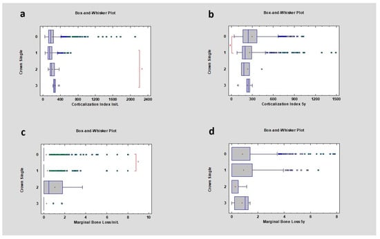
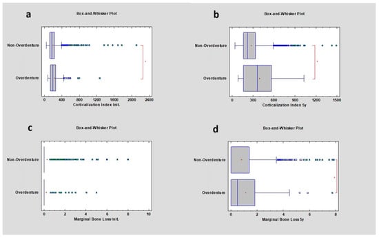
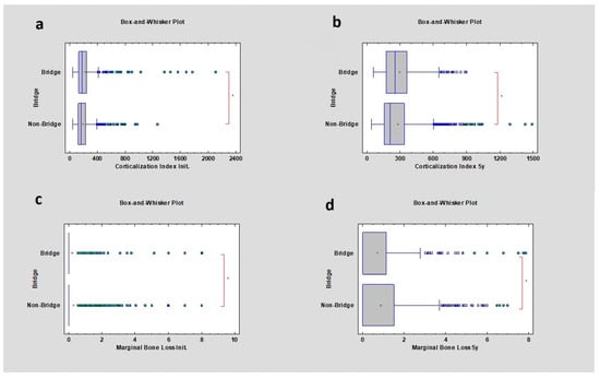
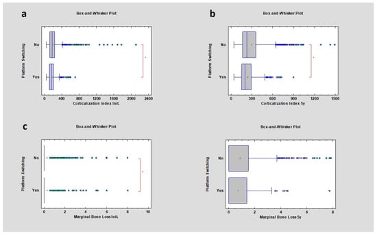
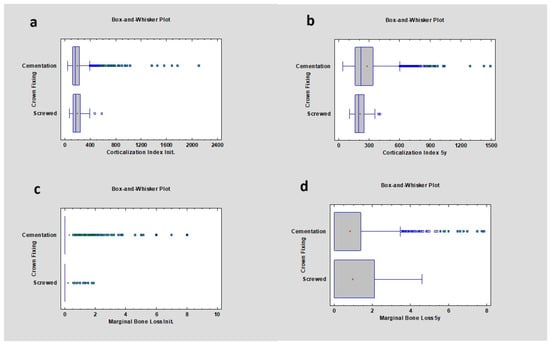
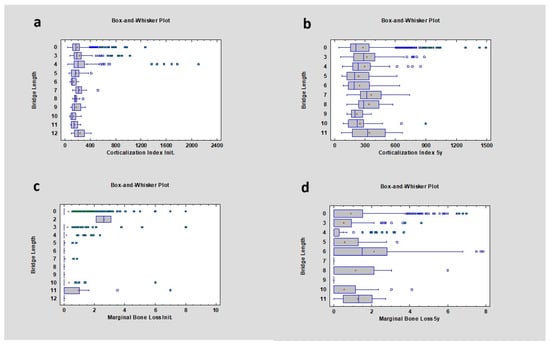
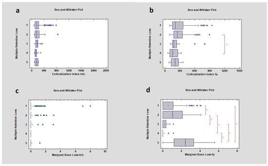
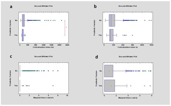
The new Radiological Corticalization Index (CI) is an indicator that describes bone remodeling near the dental implant’s neck at the pixel level and is not visible to the naked eye. The aim of this research was to evaluate the correlation between the CI and bone remodeling using only radiographic (RTG) images. RTG samples were divided into groups depending on prosthetic restoration; the implant neck area around dental implants was examined, and texture features of the RTG images were analyzed. The study also investigated the type of prosthetic restoration and its influence as a factor on bone structure. The statistical analysis included evaluating feature distribution, comparing means (t-test) or medians (W-test), and performing a regression analysis and one-way analysis of variance or the Kruskal–Wallis test, as no normal distribution or between-group variance was indicated for the significant differences in the investigated groups. Differences or relationships were considered statistically significant at p < 0.05. The research revealed correlations between single crowns, overdenture restoration, bridge restoration, platform switching, prosthetic fracture, CI, and also marginal bone loss where p was lower than 0.05. However, the corticalization phenomenon itself has not yet been fully explored. The findings suggest that, depending on the type of prosthetic restoration, the corticalization index may correlate with marginal bone loss or not. Further research is necessary, as the index is suspected to not be homogeneous.
Full article
(This article belongs to the Section Medical Imaging and Theranostics)
►
Show Figures
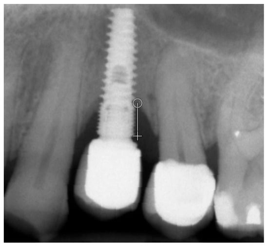
Figure 1


